
Dignity Health Sequoia
Orthopedic Surgery & Sports Medicine - Dignity Health Medical Group - Redwood City, CA
Address
Hours
Mon
Tue
Wed
Thu
Fri
Sat Closed
Sun Closed
Complete sports medicine and orthopedic care from head to toe.
The entire Dignity Health Medical Foundation team, from primary care to our orthopedic experts to neurological team, works together to treat your injuries. From personalized treatment plans to non-operative procedures, our specialists are focused on your healing and recovery from start to finish, so you can get back to all the daily activities you love.
Orthopedic conditions treated
Dignity Health orthopedic specialists have your back when it comes to specialized orthopedic care. Known for their compassion and excellence in patient care, our orthopedic specialists will help diagnose and manage conditions, relieve pain, and see you through rehabilitation.
At Dignity Health, our hospitals are staffed by teams of specialists who provide comprehensive orthopedic care. Here are some of the conditions our orthopedic specialists treat:
- Arthritis
- Bursitis
- Fibromyalgia
- Fractures
- Osteoporosis
- Hip conditions and procedures
- Scoliosis
- Low back pain
- Hand pain
- Knee pain
- Shoulder pain
- Neck pain
- Arm and elbow pain
- Foot and ankle pain
Meet our orthopedic team
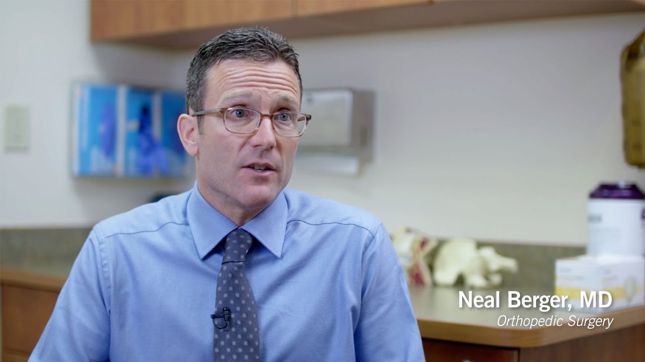
Meet Neal Berger, MD

Dr. Berger attended the University of Pennsylvania for his undergraduate education. He received his medical degree from the University of California – Davis where he was elected to the Alpha Omega Alpha (AOA) honor society. He did his orthopedic surgery residency at UCLA (University of California – Los Angeles) where he was elected as one of the Chief Residents in his graduating class. He did a sports medicine fellowship with SOAR, during which time he worked as an assistant team physician for the San Francisco Giants.
Meet Arati M. Dunbar, MD

Dignity Health Medical Group Saint Francis/St. Mary's Dr. Arati Mallik Dunbar is a unique, patient focused orthopedic surgeon with a special interest in sports medicine and arthroscopy. She is board certified in orthopedic surgery and fellowship-trained in sports medicine. Her medical practice includes both general orthopedic care and sports related injuries to shoulders, knees, elbows and ankles. She was fellowship trained at UCLA Medical Center where treatment of injured athletes was a primary focus. An athlete herself, Dr. Dunbar draws on this first hand knowledge in her diagnosis and treatment of her patients

Learn more about orthopedics
- What Is Arthroscopy?
- What is a Rotator Cuff Repair?
- What is ACL Reconstruction?
- What is Open Repair of Ankle Fractures?
- About Wrist Fractures
- What is Carpal Tunnel Syndrome?
- What is "Trigger Finger" Release?
This is a surgery done through tiny incisions ("poke holes") about ½” each. A tiny telescope-like device attached to a camera is inserted and allows the physician to see the inside of joints. The arthroscopy allows the physician to see a magnification up to 40 times. Small biters, shavers, and other tools can be use to clean up or repair tears in menisci, ligaments, etc. Using arthroscopy, the physician can often see more detail than with open surgery. The small incisions through muscle causes less pain than larger ones and provides a quicker rehabilitation after surgery.
The rotator cuff (see above) is reattached to the upper arm bone (the humerus). This is most often done with stitches attached to plastic, or metal tacks, or screws. The tacks or screws are placed in the bone and the stitch is placed through the torn part of the rotator cuff so the rotator cuff is now attached again to the bone. Patients with this condition are usually in a sling for about 4 weeks and cannot do any lifting for about 6 weeks.
A variety of tissues/structures can be used to reconstruct/replace the ACL. The most common are the patellar tendon in the front of knee and the hamstring tendons in the back of the knee. Your own tendon (autograft) or one from a cadaver (allograft) can be used. Your body essentially uses these tendons as "scaffolds" to show it where to "grow" a new ligament. First, blood vessels grow in and then new ligament cells follow. That is why when autograft is used, the new "ligament" is actually the weakest from 4 to 6 months after surgery, when the blood vessels and the cells are still "growing." When an allograft is used, it takes longer for blood vessels and the cells to start growing- sometimes years. Usually, a long leg hinged brace is used for the first 6 weeks and crutches for the first 2 to 4 weeks. Physical therapy is an important part of the recovery process and is started immediately. Dr. Dunbar typically allows patients to start a running program around 4 months after surgery, start light sports at 6 months, and return to full sports around 8 to 9 months after surgery.
There are three malleoli in the ankle, which are the prominences (or bumps) you see on the inside and outside of the ankle. The one on the inside is the medial malleolus which is part of the tibia or shin bone, while the one on the outside is the lateral malleolus and is part of the fibula or smaller bone in the leg. The posterior malleolus is a bump on the back of the tibia or shinbone but you cannot see it from the outside.
Ankle fractures almost always involve the ankle joint. If the bone is out of place and heals without the joint surface being smooth, it can act like sandpaper and wear away the cartilage, thus leading to arthritis. That is why surgery is often done for ankle fractures. A thin plate and screws are often used on the lateral malleolus (fibula), and screws and pins are usually used to fix the medial malleolus. The posterior malleolus may or may not require a screw to fix it. After surgery you may be in a splint, a cast or a fracture boot.
One of the other advantages of surgery for an ankle fracture is that you can usually start moving the ankle sooner. This may help prevent stiffness during the recovery process.
Wrist fractures can be treated in a variety of ways depending on whether they are "in place" or "out of place," and whether they involve the joint. Fractures that are completely "in place" are usually treated with a cast or brace. Fractures that are "out of place" or involve the joint may require manipulating the bone back into place. Successfully holding the bone in place may require a cast, pins, plate and screws or an external fixator. An external fixator is a device in which pins are placed through the skin and through the bone at places away from the fracture. Bars connect the pins and keep traction across the fracture which keeps the bone from collapsing back out of place. While this may look odd or scary, it actually helps in the same way as if the doctor were walking around with you trying to keep the fracture in place while holding the bone.
In this procedure, the top of the tunnel is opened up to release the pressure on the tendon and nerve. This allows the nerve to shrink back to its normal size. Depending on how much damage has already been done to the nerve, you may or may not have all of your symptoms go away. Pain usually goes away right away, but full feeling and strength may never return. The surgery is done as an outpatient and often with just a local anesthetic.
In this surgery the pulley is cut and allowed to heal in a more opened-up position like a "drawbridge." (This is also like letting out the waistband of those shrunken jeans!) This is usually done as an outpatient procedure and often with a local anesthetic.
- What is Impingement Syndrome?
- What is a Rotator Cuff Tear?
- What is an Anterior Cruciate Ligament Tear?
- What is a Dislocated Shoulder?
- What is a Separated Shoulder?
- What is Carpal Tunnel Syndrome?
- What is "Trigger Finger"?
This used to be called Bursitis in the shoulder. There is a bursa on top of the rotator cuff and underneath the flat bone (acromion). When the rotator cuff is irritated, the body's natural reaction is to try and cushion it by filling the bursa with fluid. This in turn puts more pressure on the rotator cuff. When the rotator cuff gets more irritated it can scar or tear, leading to more pain, or more weakness.
This is a tearing of one or more tendons of the rotator cuff. It usually tears where it is attached on the arm. It can be caused by one major trauma, or over time in patients that have Impingement Syndrome (see definition above).
The anterior cruciate ligament is one of four major ligaments that connect the femur (thigh bone) and tibia (shin bone) and keeps the tibia from moving too far forward relative to the femur. When you tear your ACL (anterior cruciate ligament) in your knee, it has to be reconstructed, not repaired. This is because there is not a very good blood supply to the center of the ligament, and simply stitching the tear is not adequate. The most common ways to tear the ACL is with hyperextending the knee, as when you catch the tip of ski and your knee bends the wrong way. In football, a helmet to the outside of the knee, a so called valgus injury, may result in the terrible triad: an ACL tear, a medial collateral ligament (MCL) tear – the ligament on the inside of the knee, and a tear of the medial meniscus – the cartilage cushion on the inside portion of the knee. The ACL can be tear w/ other mechanisms but these are the more common ways. The ACL is important in pivoting or cutting. That is why patients who tear their ACL can often perform straight ahead type activities like running or weightlifting but are unable to play football, basketball, or soccer.
This is where the "ball" (humeral head) part of the joint comes out of the "socket" (glenoid) portion of the joint. The shoulder is a generally unstable joint. It is like a big ball sitting on a small flat saucer. This gives you the range of motion you have in your shoulder.
By comparison, in the hip, the ball sits in the socket, but most people don't have the same range of motion in their hips as they do in their shoulders.
There are two main static stabilizers in the shoulder. The labrum is a "rubbery" substance like a meniscus that goes around the socket and acts like a rubber stop. The capsule or joint lining is a sac that surrounds the joint. The dynamic stabilizer (which can strengthen or relax) is the rotator cuff. When you dislocate your shoulder, the structures that can be injured are the labrum, the capsule, the humeral head and/or the glenoid. In older patients, the rotator cuff may also be injured.
The shoulder usually dislocates in the front, but can also come out the back, typically in patients with seizures.
This involves the AC joint, which is between the collarbone (clavicle) and the flat part on the top of shoulder blade (acromion). When the ligaments in this joint tear, you then have a separated shoulder. It may first appear that the collarbone is pushing up, but actually the shoulder blade has dropped.
This is a big "tunnel" on the palm side of your wrist. There are eight tendons inside along with the median nerve. The median nerve supplies feeling to the thumb, the index finger, the middle finger and usually half of the ring finger. It also supplies the "power" to most of the muscles which move the thumb. When the tendons become irritated, they become swollen. This can occur, for example, when you are typing on a computer for a long period of time. Because the tunnel is a fixed size, when there is swelling, there is less room for the nerve and it becomes pinched. It is similar to your gaining five pounds and shrinking your jeans-either the inside has to get smaller or the outside has to get bigger.
The way to make the inside smaller is to get rid of the irritation in the tendons and shrink the tendons back to their normal size. This can be done with rest, braces, medications and/or injections. If the nerve is pinched for long periods of time, then in addition to the pain, there can be tingling, numbness or even weakness. When the symptoms become more severe, your Doctor may have you get an EMG, which tests the function of the nerve. If there is too much pinching of the nerve or you have not gotten better, he or she may recommend a Carpel Tunnel Release (see below).
Tendons that bend the knuckles of the finger start in the forearm. Because they are so long, they have arches or pulleys along the way to help pull them close to the bone. One of the arches, the A1 Pulley, is located where the finger connects to the palm. Because this is the area where there is a lot of movement of the tendon, there can be irritation. When the tendon gets irritated, it swells and rubs on the pulley. When the pulley gets irritated, it scars or shrinks (tightens). Because of this it "catches" and that is where term "trigger finger" comes from. The finger can even get stuck when the swollen portion moves past the pulley and can't pull back. Again, this is like gaining five pounds and shrinking your jeans. You now either have to make the inside smaller or the outside bigger.
You can sometimes make the inside smaller with rest or an injection. If this doesn't work, you may require a trigger finger release (see above).
Unless you're a physician yourself, orthopedic definitions can be overwhelming. Here is a list of relatively simple explanations to help you better understand the human body and its many common orthopedic structures and conditions:
What is a Ligament?
This is a "rope-like" structure that connects one bone to another bone and keeps the bone from going in the wrong direction.
What is a Tendon?
This is a "rope-like" structure that connects the muscle to the bone. The muscle pulls on the tendon, the tendon pulls on the bone and the bone moves.
What is Cartilage?
Articular cartilage is the white, firm, gliding surface on the ends of bones and on the back of knee caps. It looks like the end of a chicken bone. It helps the joints glide smoothly. When the articular cartilage is worn away, the result is arthritis.What is Meniscus?
Meniscus is made up of fibrocartilage. It looks and feels "rubbery" and acts like a shock absorber. You have two menisci in each knee and one in the joint between the collarbone and shoulder blade.
What is a Joint?
A joint is the area where two bones come together and move independently Most joints are synovial, which means they have a joint lining and fluid inside and have articular cartilage on the end to help the joint move and glide.
What are Membranous Joints?
This is another type of joint that has no fluid or joint lining. An example is the connecting points between the sternum (breast bone) and ribs.
What is Bone Actually Made Of?
Bone is the body's framework that is made of cells, calcium and other minerals. There is marrow in the center, which makes the red blood cells that carry oxygen. As the body ages, the marrow in some bones begins to contain a higher fat content.What is a Rotator Cuff?
The shoulder is an inherently unstable joint. The rotator cuff helps keep the shoulder in place. It consists of four muscles and their four tendons (see definitions above). Two are in the back, one is on top, one is in the front. They are small muscles, and they are not strong, but they have a lot of endurance.What is a Bursa?
This is a thin lubricating sac that can get bigger when it is trying to "cushion" areas of irritation. Unfortunately, the fluid that your body uses to fill it up can actually cause more irritation and pain than that experienced with the initial problem.What is a Fracture?
A fracture is any break in a bone that goes partially or fully through the bone. During a fracture, the bone can remain in place or it can move out of place.What is Dislocation?
Dislocation always involves a joint, as when one bone moves out of joint relative to the other bone.What is a Sprain?
This is any stretching, partial tearing or complete tearing of a ligament (see definition above).What is a Strain?
This is any stretching or tearing of the muscleWhat is a Rupture?
This is when a tendon pulls off from its attachment to a bone.
Care when you need it
With clinics throughout California, a Dignity Health Medical Foundation physician is right around the corner, waiting to assist you along your wellness journey.

Find a Doctor
Looking for a doctor? Perform a quick search by name or browse by specialty.